Imaging at the speed of life
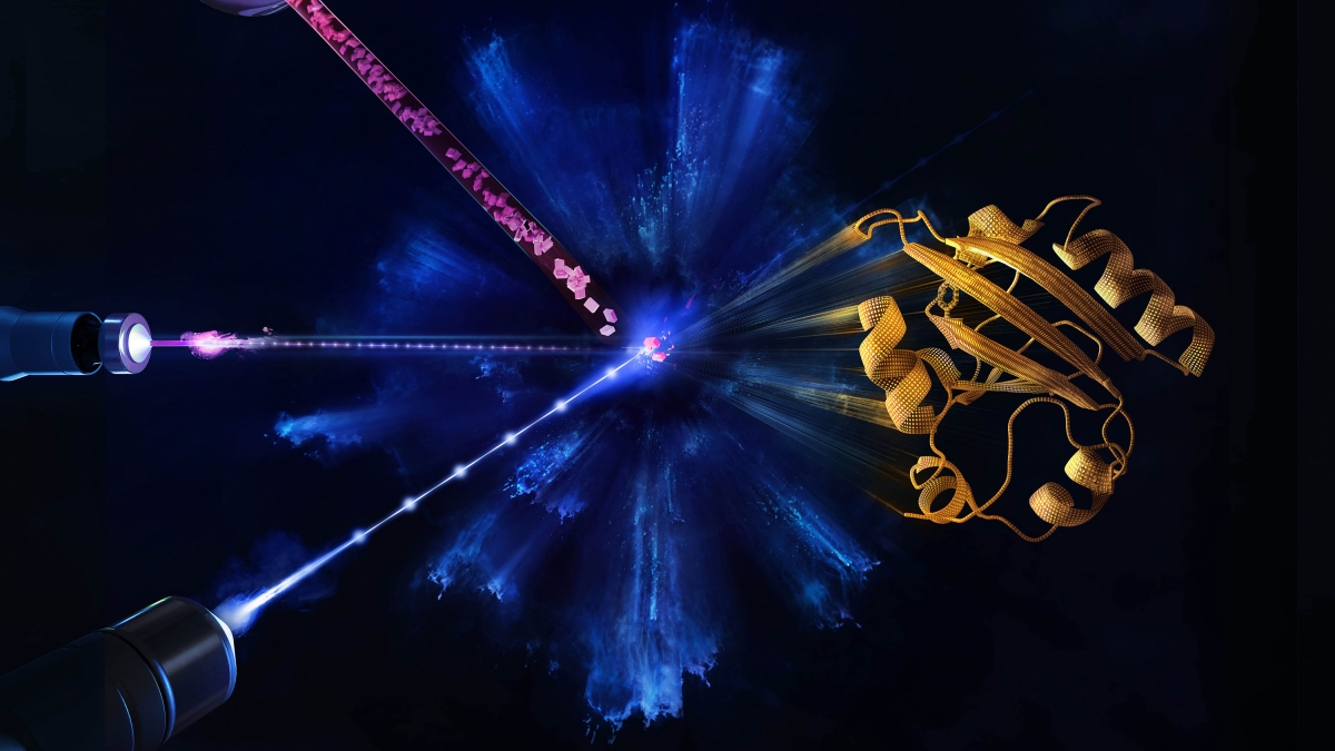
The EuXFEL produces intense X-rays in extremely short pulses at a rate of one megahertz — a million pulses a second. The rays are aimed at crystals containing proteins, in a method called X-ray crystallography. When a crystal is hit by the X-ray pulse, it diffracts the beam, scattering in a certain pattern that reveals where the atoms are and producing a “snapshot.” Copyright: European XFEL / Blue Clay Studios
To study the swiftness of biology — the protein chemistry behind every life function — scientists need to see molecules changing and interacting in unimaginably rapid time increments: trillionths of a second or shorter.
Imaging equipment with that kind of speed was finally tested last year at the European X-ray Free-Electron Laser, or EuXFEL.
Now, Arizona State University researchers join a team of physicists from the University of Wisconsin-Milwaukee and their international collaborators to present EuXFEL’s first molecular movie, or “mapping,” of the ultrafast movement of proteins.
With this capability, scientists can watch how proteins do their jobs properly — or how their shape-changing goes awry, causing disease.
“Creating maps of a protein’s physical functioning opens the door to answering much bigger biological questions,” said Marius Schmidt, a professor of physics at the University of Wisconsin-Milwaukee (UWM) who designed the experiment and is lead author of the new study. “You could say that the EuXFEL can now be looked on as a tool that helps to save lives.”
Petra Fromme, who directs the Biodesign Center for Applied Structural Discovery, is very excited about the impact of the study.
"All processes in nature are highly dynamic, but most structures determined to date show only a static picture of a molecule. Time-resolved serial femtosecond crystallography at the European XFEL allows us to 'see,' in atomic detail, the dynamics of this photosensor protein in real time," Fromme said.
Fromme helped oversee ASU’s contribution to the multifaceted EuXFEL project. In addition to her research at the Biodesign Institute, she is a Regents Professor in ASU’s School of Molecular Sciences.
The group’s findings signal the arrival of a new age of protein research enabling enzymes involved in disease to be observed in real time for meaningful durations in unprecedented clarity. The paper is published online in the journal Nature Methods.
Blaze of light
The EuXFEL produces intense X-rays in extremely short pulses at a megahertz rate — a million pulses a second. The rays are aimed at crystals containing proteins, in a method called X-ray crystallography. When a crystal is hit by the X-ray pulse, it diffracts the beam, scattering in a certain pattern that reveals where the atoms are and producing a “snapshot.”
The rapid-fire X-ray pulses produce 2D snapshots of each pattern from hundreds of thousands of angles where the beam lands on the crystal. Those are mathematically reconstructed into moving 3D images that show changes in the arrangement of atoms over time.
The European XFEL, which opened last year, has taken this atom mapping to a new level. Extremely powerful bursts contain X-ray pulses at a quadrillionth of a second, in “bursts” that occur at 100 millisecond intervals.
The new experiments began with a flash of blue, visible light that induced a chemical reaction inside the protein crystal, followed immediately by a burst of intense X-rays in megahertz pulses that produce the “snapshots.”
Initial research was staged in 2014 at the U.S. Department of Energy’s SLAC National Accelerator Laboratory in California, where Fromme, Schmidt and their colleagues were able to document atomic changes in their protein samples for the first time at an XFEL.
Subsequently, in 2016, they were able to map the rearrangement of atoms in the range of time proteins take to change their shapes — quadrillionths of a second (femtoseconds) up to 3 trillionths of a second (picoseconds). In a picosecond, which is a trillionth of a second, light travels the length of the period at the end of this sentence.
Previous time-resolved crystallography on their photoreactive protein had already been completed using other X-ray sources capable of imaging time scales larger than 100 picoseconds, leaving a gap of uncharted time between 3 and 100 picoseconds that the scientists were able to fill using the EuXFEL.
The exceptional brightness of the laser and the megahertz X-ray pulse rate allows much quicker data gathering with greater resolution over longer time frames.
Europe’s X-ray super laser
The EuXFEL is an extreme device. At 3 kilometers long, spanning the distance between the German federal states of Hamburg and Schleswig-Holstein, it is the largest XFEL in the world. Superconductive technology is used to accelerate high-energy electrons, which generates the X-rays.
For their experiments, the scientists used the instrument for Single Particles, Clusters, and Biomolecules and Serial Femtosecond Crystallography (SPB/SFX) at EuXFEL. The instrument is designed to use the extremely short X-ray pulses for imaging and crystallography experiments in order to explore the structure and dynamics of small biological samples.
The team at the SPB/SFX instrument are able to adjust the parameters for each experiment depending on the protein being studied. These include changing the spot size of the optical light pulse to take into account the size of the individual protein crystals, and the time delay between optical and X-ray laser to accommodate the duration of the reaction being studied.
“The knowledge and expertise we have acquired with these experiments at the European XFEL will enable scientists to use the SPB/SFX instrument to produce molecular movies of their sample,” said Adrian Mancuso, European XFEL group leader at the SPB/SFX instrument where the study was done.
The new study extends recent research, promising important medical advances by means of enhanced crystallography with XFELs. Using this method, Fromme and fellow researchers have witnessed how multiple proteins work together, how enzymes responsible for antibiotic resistance disable a drug and how proteins change their shape in order to absorb light and enable sight.
The first author of the new study is doctoral student Suraj Pandey, who came to UWM from his native Nepal. Pandey’s role was to analyze the data and calculate the maps of structural change. Of the millions of X-ray pulses that XFELs deliver, the majority don’t hit a target at all. In fact, only 1% to 2% diffract off a protein crystal, while the remaining pulses produce “noise” that must be removed from the data.
Growing the specialized protein crystals required for the experiments was also challenging, and Pandey says that some of the invaluable specimens melted during their transit to Germany when they were held up at customs.
The ultimate success of the new experiments gave Pandey tremendous satisfaction.
“It’s a one-of-a-kind technology,” he said of the EuXFEL. “We pioneered the usage of European XFEL in seeing the movies of how proteins function. I’m just flying.”
Molecular cinema
Traditional structural biology methods use X-rays to produce snapshots of the 3D structure of molecules, including proteins. Although valuable, this information does not reveal details about the dynamics of biomolecular processes. If several snapshots can be taken in fast enough succession, however, these can be pasted together to make a so-called molecular movie.
The high repetition rate of the extremely short X-ray pulses produced by the European XFEL makes it now possible to collect large amounts of data to produce movies with more frames than ever before. An international group of scientists have now worked out how to make optimal use of the European XFEL’s very high X-ray repetition rate to make these molecular movies at the facility in order to reveal unprecedented details of our world.
Schmidt is also enthusiastic about the power of XFEL technology to revolutionize scientist’s understanding of dynamical properties that had long remained out of reach for investigation.
“Imagine a movie of a downhill ski race made up of only three frames. This will tell you something about how the race began and finished, and give you an image of the skier as well as a glimpse of the landscape somewhere on the mountain, but not much else. A movie containing many more frames, however, would follow the race in detail and would be much more informative,” he said. “Now that we understand how to harness the European XFEL X-ray pulses to collect large amounts of data to produce detailed molecular movies, we are very excited to see what unimagined insights are now possible.”
The work was carried out with BioXFEL collaborators from Arizona State University and the Deutsches Elektronen-Synchrotron (DESY) and beamline research specialists at the EuXFEL.
ASU coauthors on the paper include members of the Biodesign Center for Applied Structural Discovery and ASU’s School of Molecular Sciences including Jorvani Cruz Villarreal, Austin Echelmeier, Diandra Doppler, Raimund Fromme, Daihyun Kim, Alexandra Ros, Petra Fromme as well as Stella Lisova from ASU’s Department of Physics.
More Science and technology

Compact X-ray laser lab aims to reveal deep secrets of life, matter and energy
X-rays allow us to view inside the human body to diagnose broken bones and other hidden problems. More recent X-ray advances are…

Apollo lunar samples enable ASU researcher to pinpoint moon’s crystallization timeline
A team of researchers, including Arizona State University geochemist Melanie Barboni, in collaboration with scientists from The…

NASA launches space telescope to chart the sky and millions of galaxies
California’s Vandenberg Space Force Base was the site for Tuesday’s 8:10 p.m. launch of the NASA SPHEREx mission aboard a SpaceX…