One of the current paradigms in cancer treatment is not to treat a tumor itself. Rather, therapeutics can focus on a tumor’s microenvironment — the area where tumor cells and a patient’s healthy tissues collide.
Mehdi Nikkhah, an assistant professor of biomedical engineering in the Ira A. Fulton Schools of Engineering at Arizona State University, has been working for the past five years on bioengineering a way to study the tumor microenvironment.
In a project led by recent ASU biomedical engineering doctoral graduate Danh Truong, a multidisciplinary team made a discovery of a new role that fibroblast cells play in the spread of breast cancer tumors using microfluidic tumor models. The results were recently published in a highly influential research journal, Cancer Research, after rigorous peer review.
Truong, who is now conducting postdoctoral research at MD Anderson Cancer Center in Houston, says he is proud to have published in the American Association for Cancer Research’s reputable journal.
“Many of the researchers that I admire have all published in this journal,” Truong said. “It is definitely a proud moment when I realize our research stands alongside theirs.”
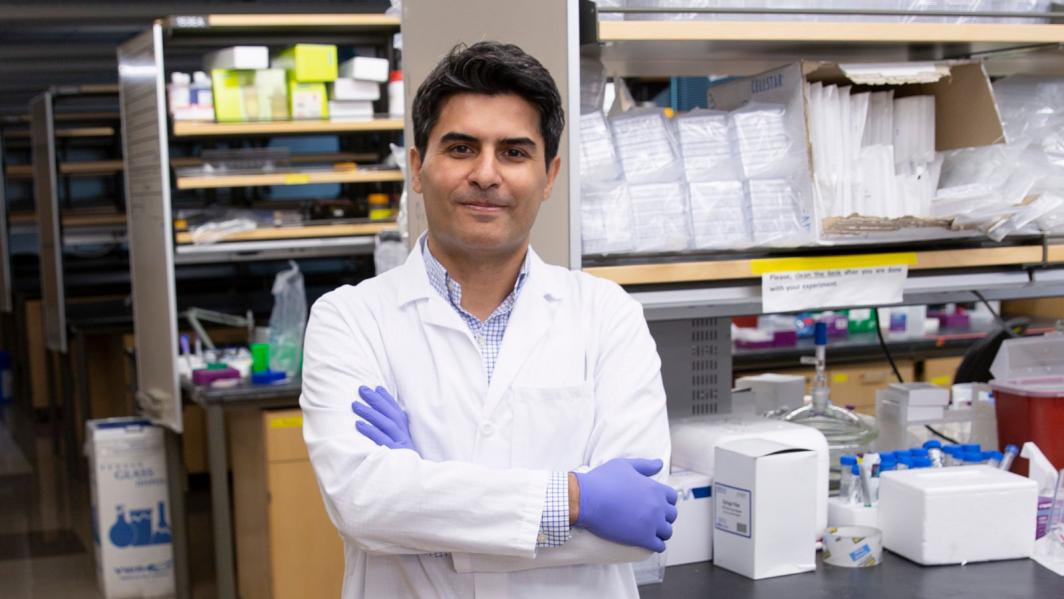
Assistant Professor Mehdi Nikkhah in his lab, where he and his students study the interface of micro/nanotechnology, advanced biomaterials and biology. Photo by Erika Gronek/ASU Now
Isolating the tumor microenvironment on a chip
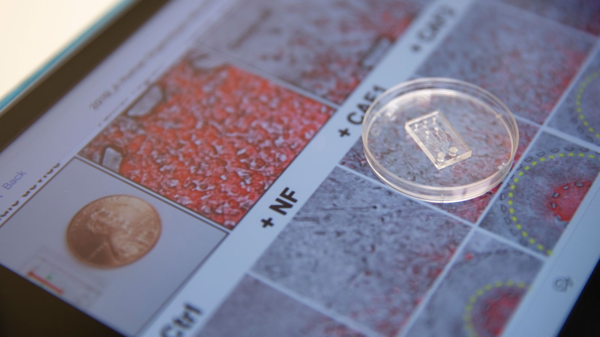
Nikkhah, Truong’s primary doctoral studies adviser, uses microengineered microfluidic chips — rectangular pieces of plastic about the size of a long fingernail with specially designed channels to deposit live cells — to replicate disease states or organs in an easily controlled environment. Depending on the biological feature being studied, these microfluidic chips are often called “organ on a chip” or “disease on a chip.” For this project, Nikkhah and the research team created breast cancer on a chip.
“We can replicate a patient’s specific tumor on this chip and manipulate and control the environment precisely,” said Nikkhah, a faculty member in the School of Biological and Health Systems Engineering, one of the six Fulton Schools.
Such chips can one day replace animal models, such as mice, which scientists have historically used as hosts to study cancerous tumors. However, animal biology is different than human biology, and many complicating factors arise when trying to study cancer cells in these environments. These on-chip models also replace 2D methods of culturing tumor cells on plate assays, which do not accurately replicate a human’s surrounding tissues.
By using the 3D design of a chip that in part replicates a human’s tumor microenvironment, Nikkhah and the research team are able to examine exactly what fibroblast cells — which make up the connective tissue that assists in wound healing — do to promote tumor progression.
“(This model) can mimic, to an extent, the same interaction that happens in the human body, where cancer-associated fibroblasts have been shown to enhance cancer invasion,” Truong said.
The team collected fibroblast cells from biopsy samples of three Mayo Clinic breast cancer patients with the help of Mayo Clinic surgical oncologist Barbara Pockaj and obtained commercially available cancer cells of the same subtypes to replicate the patient tumors. This allowed the researchers to better understand the role of fibroblasts in breast cancer metastasis, or when tumors spread and create secondary tumors elsewhere in the body.
Through high-resolution imaging, the team can see individual cancer cells and fibroblast cells, and measure the speed of a single cancer cell moving in the engineered microenvironment. This phenomenon would otherwise be impossible to observe in a person’s body or an animal model.
Watching good cells break bad
The research team observed cancer cells speeding up when they came into contact with fibroblast cells.
Using a technique called RNA sequencing, which revealed thousands of possible molecular targets (conventional methods reveal less than 100 targets), the team made a novel discovery.
The team identified a gene called GPNMB that appeared only in samples from patients with highly aggressive triple-negative breast cancer. In publicly available clinical data tracking nearly 3,000 patients, those with high expression of the GPNMB gene had decreased survival rates.
“Our big ‘aha’ moment happened when we were poring over the data and comparing what we found to literature data,” Truong said. “We uncovered a possible target, the protein GPNMB, which was corroborated with data in literature, but not yet observed in the interaction between cancer cells and cancer-associated fibroblasts. We thought this protein may be involved in the interaction and decided to disrupt it.”
When they silenced that gene’s expression in cancer cells in the chip model, the cancer cells stopped spreading to the surrounding tissue.
“The microenvironment is inducing changes in gene expression of cancer cells,” Nikkhah said, “specifically because of fibroblast cells that lead them to be in a highly invasive state.”
In a typical wound, like a cut on your hand, fibroblasts migrate to the wound and proliferate to heal the cut. But when they encounter a “wound” of cancer cells, the cancer cells use molecular signaling to turn fibroblasts into their helpers to accelerate the growth and invasion of cancer cells.
“The fibroblasts change their phenotype in the tumor microenvironment in a way to help the tumor spread,” Nikkhah said.
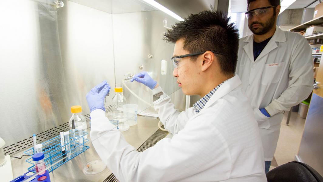
Danh Truong, a recent ASU biomedical engineering doctoral graduate now conducting postdoctoral research at MD Anderson Cancer Center in Houston, works in Mehdi Nikkhah’s lab. Truong led the project as part of his dissertation research. Photo by Nora Skrodenis/ASU
Engineers and biologists collaborate to generate new knowledge
Cancer research is a multidisciplinary field that requires more than bioengineering expertise. In addition to working with Mayo Clinic oncologists and pathologists, Nikkhah and Truong collaborated with the ASU Biodesign Institute to accomplish gene expression analysis and bioinformatics.
Joshua LaBaer, professor, executive director of the ASU Biodesign Institute and cancer biologist, provided input on what cell types the research team should use and input on the biology behind replicating a patient’s tumor on a chip.
Bioinformatics scientist and ASU Biodesign Institute Assistant Research Professor Jin Park analyzed gene expression level data obtained by RNA sequencing.
“As the data are complex, we need specialized bioinformatics and biostatistics to process and analyze them to discover important genes regulating cancer invasion and draw biological conclusions on the gene functions,” Park said.
The interdisciplinary partnership between the School of Biological and Health Systems Engineering, one of the six Fulton Schools, and the ASU Biodesign Institute helps bioengineers address many challenging scientific questions, such as finding cures for cancer.
“This paper is a prime example of what we can achieve through such collaboration, where cell biology, bioinformatics and microfluidics techniques were synergistically utilized to identify novel cellular interactions and gene functions that may guide future development of novel therapies for metastatic breast cancer,” Park said.
The team’s collaboration also extended to the University of Arizona Cancer Center, where Ghassan Mouneimine, assistant professor and cancer biologist, gave the team input on cell migration experiments.
Beyond the multidisciplinary nature of the project, the research was conducted by a multilevel team comprised of undergraduate students and doctoral students as well as junior and senior faculty. Involving undergraduates early on in their academic careers helps students find opportunities to mature in their scientific careers and build personal and professional skills, Nikkhah said.
“These highly talented and motivated students are at the beginning of their path to become a true force of change in the world and make a positive impact on society,” Nikkhah said. “Our role as educators is to guide them to step into the right path, discover their talents and to get to know their strengths and weaknesses.”
As a doctoral student, Truong also learned valuable skills that contributed to his current position at one of the top cancer centers in the country.
“The incorporation of graduate and undergraduate students in research is definitely a positive thing,” Truong said. “Research labs are environments where students can tinker and learn while being protected from the real-world consequences of failures. I cannot recall how many times I have failed, but each time gave me the foresight and experience for my next experiment, which eventually led to my success. Without my research experiences in Dr. Nikkhah’s lab, I would not be where I am today.”
A step toward better personalized medicine for cancer treatment
The research this team conducted will lead to new ways to study therapeutics and create personalized medicine for each patient’s particular cancer and tumor microenvironment.
“If you can understand how cancer spreads, you can design better strategies to stop it,” Nikkhah said.
Once scientists can replicate a person’s specific tumor and its surrounding microenvironment — including immune cells and other proteins and tissues — on a chip, they can add drugs to see how the cancer responds without having to test them on a patient or incompatible animal model.
The chips are also inexpensive, so hundreds of tests can be run at low cost compared to tests on animal models. In addition to being more cost-effective, the chips are more scientifically effective and improve greatly on the low success rate of animal model clinical trials.
“It’s not very far off that we can create fully patient-derived tumor and tissue cells and potentially go to fully personalized medicine for cancer therapy,” Nikkhah said. “If you design the therapeutics to knock down targeted genes, for instance GPNMB, inside the patient’s tumor cells, you may be able to stop the spread of the tumor.”
The microfluidic chip can be used on a number of different cancer types. For example, Nikkhah has worked with Banner Neurological Institute (BNI) to create a glioblastoma brain tumor on a chip. Each cancer’s microenvironment requires a slightly different chip design, but there are commonalities among all cancers that make the chip a viable option for study.
The progress the research team has made is reinforced by their work’s publication in Cancer Research.
“As an institution without a medical school, this paper will certainly help boost the status of ASU toward a major research university in cancer research,” Park said.
Preparing to break new ground on cancer research
After spending the past five years developing the chip and learning the biology behind fibroblasts’ role in cancer metastasis, Nikkhah is looking to isolate all cell types derived from a single patient to study on the chip. This would help to better cross-correlate their findings with the same patient's data or other clinical data. A fully patient-derived cancer on a chip model would be a big step toward using the chips for personalized medicine.
Nikkhah also recently earned a second National Science Foundation grant to build on the past four years’ work that resulted in the Cancer Research paper. Next, he’ll be studying the mechanism of anticancer drug resistance using the same chip platform.
“The chip has shown promise,” Nikkhah said, “so the NSF wants us to continue this work to study the mechanisms of drug resistance to help design better therapeutics.”
Top photo: A microfluidic chip created by a team of Arizona State University researchers sits on top of images of cancer cells and fibroblast cells. Results of research involving studies of the cells were recently published in the American Association for Cancer Research’s (AACR) Cancer Research journal. The project was led by recent ASU biomedical engineering doctoral graduate Danh Truong and Assistant Professor Mehdi Nikkhah, a faculty member in the School of Biological and Health Systems Engineering, one of the six Ira A. Fulton Schools of Engineering at ASU. The bioengineers collaborated across disciplines with researchers in the ASU Biodesign Institute, Mayo Clinic Arizona and the University of Arizona Cancer Center. Photo by Erika Gronek/ASU
More Science and technology

Applied Materials invests in ASU to advance technology for a brighter future
For nearly 60 years, global giant Applied Materials has been hard at work engineering technology that continues to change how microchips are made.Their products power everything from flat-panel…

Meet ASU engineering students who are improving health care, computing and more
Furthering knowledge of water resource management, increasing the efficiency of manufacturing point-of-care health diagnostic tools and exploring new uses for emerging computer memory are just some…

Turning up the light: Plants, semiconductors and fuel production
What can plants and semiconductors teach us about fuel production?ASU's Gary Moore hopes to find out.With the aim of learning how to create viable alternatives to fossil-based fuels, Moore — an…


