Researchers discover regenerated lizard tails are different from originals

Just because a lizard can grow back its tail, doesn’t mean it will be exactly the same. A multidisciplinary team of scientists from Arizona State University and the University of Arizona examined the anatomical and microscopic makeup of regenerated lizard tails and discovered that the new tails are quite different from the original ones.
The findings are published in a pair of articles featured in a special October edition of the journal The Anatomical Record.
“The regenerated lizard tail is not a perfect replica,” said Rebecca Fisher, an associate professor in ASU’s School of Life Sciences, and at the UA College of Medicine – Phoenix. “There are key anatomical differences including the presence of a cartilaginous rod and elongated muscle fibers spanning the length of the regenerated tail.”
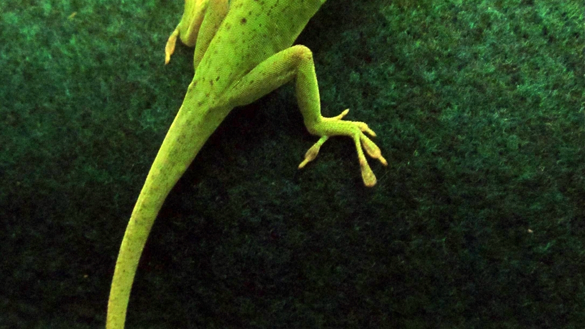
Researchers studied the regenerated tails of the green anole lizard (Anolis carolinensis), which can lose its tail when caught by a predator and then grow it back. The new tail had a single, long tube of cartilage rather than vertebrae, as in the original. Also, long muscles span the length of the regenerated tail compared to shorter muscle fibers found in the original.
"These differences suggest that the regenerated tail is less flexible, as neither the cartilage tube nor the long muscle fibers would be capable of the fine movements of the original tail, with its interlocking vertebrae and short muscle fibers," Fisher said. "The regrown tail is not simply a copy of the original, but instead is a replacement that restores some function."
While the green anole lizard’s regenerated tail is different from the original, the fact that lizards, unlike humans, can regenerate a hyaline cartilage skeleton and make brand new muscle is of continued interest to scientists who believe learning more about regeneration could be beneficial to humans in the future.
"Using next-generation technologies, we are close to unlocking the mystery of what genes are needed to regrow the lizard tail,” said Kenro Kusumi, an associate professor in School of Life Sciences in the College of Liberal Arts and Sciences, and co-author of the papers. “By supercharging these genes in human cells, it may be possible to regrow new muscle or spinal cord in the future."
“What is exciting about the morphology and histology data is that these studies lay the groundwork for understanding how new cartilage and muscle are elaborated by lizards,” said Jeanne Wilson-Rawls, co-author and associate professor in the School of Life Sciences. “The next step is understanding the molecular and cellular basis of this regeneration.”
Another interesting finding is the presence of pores in the regenerated cartilage tube. While the backbone of the original lizard tail is made of many bones with regular gaps, allowing blood vessels and nerves to pass through, in the regenerated tail, only blood vessels pass through the cartilage tube pores. This observation suggests that nerves from the original tail stump grow into the regenerated tail.
The researchers hope their findings will help lead to discoveries of new therapeutic approaches to spinal cord injuries and diseases such as arthritis.
The research team included Jeanne Wilson-Rawls, Kenro Kusumi, Alan Rawls, and Dale DeNardo from ASU’s School of Life Sciences and Rebecca Fisher from School of Life Sciences and University of Arizona College of Medicine-Phoenix.
This research was funded by Arizona Biomedical Research Commission, grant #1113, and National Center for Research Resources and the Office of Research Infrastructure Programs (ORIP) of the National Institutes of Health, grant #R21 RR031305.